
Introduction
Anaplastic lymphoma kinase (ALK) inhibitors have revolutionized the treatment of ALK-positive non-small cell lung cancer (NSCLC), significantly improving survival and quality of life. Among second-generation ALK inhibitors, alectinib has been widely used due to its superior efficacy and safety profile compared to first-generation agents like crizotinib, as demonstrated in the phase III ALEX trial1 and the J-ALEX trial in Asian population.2 More recently, alectinib has been approved for patients with resected ALK-positive NSCLC based on significant improvement of disease-free survival over adjuvant platinum-based chemotherapy.3 However, rare but severe cardiac adverse effects, such as fatal arrhythmias, pericardial effusion, or cardiac dysfunction, have been reported.4 Tako-Tsubo syndrome (TTS) is a non-ischemic and non-inflammatory cardiomyopathy often developed in postmenopausal women after exposure to physical or emotional stress. It usually curses with left ventricular dysfunction, typically reversible, and coronary syndrome or myocarditis are the most common differential diagnosis.5,6 Even though TTS has been described after several anti-tumoral drugs (cyclophosphamide, paclitaxel, doxorubicin, tyrosine-kinase inhibitors, immune-checkpoint inhibitors), it is still underreported and its pathological mechanisms poorly understood, creating challenges in identifying risk factors and optimal management strategies.7
Cancer has been reported in approximately 15-30% of patients with TTS. Compared with classic TTS, cancer-associated cases are more frequently associated with physical triggers (such as infections, invasive procedures, or treatment related complications) and present few distinct clinical features, including a higher proportion of male patients and an overall worse prognosis.8 Anticancer therapies have been increasingly recognized as potential triggers, underscoring the importance of considering TTS in oncological patients presenting with acute cardiac dysfunction.9,10
This case highlights a rare instance of stress-induced cardiomyopathy in a patient receiving alectinib and underscores the importance of vigilance and reporting uncommon cardiac toxicities. The patient provided written informed consent for the publication of this case report, including all clinical and imaging data.
Clinical case
A 75-year-old Caucasian woman with obesity and controlled essential hypertension, was diagnosed in 2014 with ALK fusion-positive, Stage IV lung adenocarcinoma with a perihilar mass in the right superior lobe with mediastinal adenopathies and bilateral lung metastases. Computed tomography scan imaging confirmed the absence of pericardial effusion and no evidence of direct invasion or significant contact between the tumor mass and the myocardial or pericardial structures. After receiving a first-line treatment with standard chemotherapy (carboplatin + pemetrexed), in 2017, the patient presented lung tumor progression, starting treatment with crizotinib until 2019. At this moment and due to lung tumor progression, a third-line treatment with alectinib at a dose of 600 mg/12 h was started in October 2019.
Two weeks later, the patient presented to the emergency department complaining of exertional shortness of breath, which had gradually worsened over the last 2 weeks. Symptoms started after a sudden syncope. In the emergency department, a diagnosis of a non-ST-segment elevation myocardial infarction was established based on the presence of pulmonary congestion, compatible electrocardiogram (EKG) (Fig. 1), and elevated cardiac biomarkers (high sensitivity Troponin-I 1820 pg/mL [normal < 17 ng/L]; NT-proBNP 2739 pg/mL [normal < 300 ng/L]). She was admitted to the coronary care unit and started on diuretics with a good response. A transthoracic echocardiogram showed a non-dilated left ventricle, with mid-apical ballooning and hyperkinesis of the basal segments. Left ventricular ejection fraction (LVEF) was 25% (Fig. 2). Coronary angiography (Fig. 3) did not demonstrate any major epicardial vessel obstruction causing acute myocardial infarction and cardiac magnetic resonance imaging (CMR) revealed myocardial edema in the hypokinetic segments, with no evidence of late gadolinium enhancement (LGE) (Fig. 4). Coronary obstruction was discarded, and although myocardial edema on CMR imaging could raise concern for alternative diagnoses such as acute myocarditis, that was considered unlikely in the absence of LGE nor previous clinical signs of infection. These findings were consistent with the Mayo Clinic criteria for the diagnosis of TTS.10 After other triggers such as pheochromocytoma (no evidenced in a recent body-CT scan), emotional trauma or pain, were absent in the clinical work-out and in her recent medical history, alectinib was considered the most likely etiology. While cancer-related stress may have contributed, the clear temporal relationship with alectinib initiation and the already known diagnosis of malignancy at this time, supported a drug-induced TTS in the absence of alternative causative causes. The event was reported in a timely manner through the Spanish Pharmacovigilance System (Agencia Española de Medicamentos y Productos Sanitarios [AEMPS]), in accordance with current pharmacovigilance recommendations.
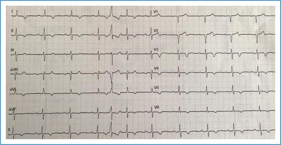
Figure 1. ECG on admission showing sinus rhythm, first atrioventricular grade block and typical ECG features of Tako-Tsubo syndrome, including residual submillimeter anterior ST-segment elevation, widespread T-wave inversion, and corrected QT interval prolongation.
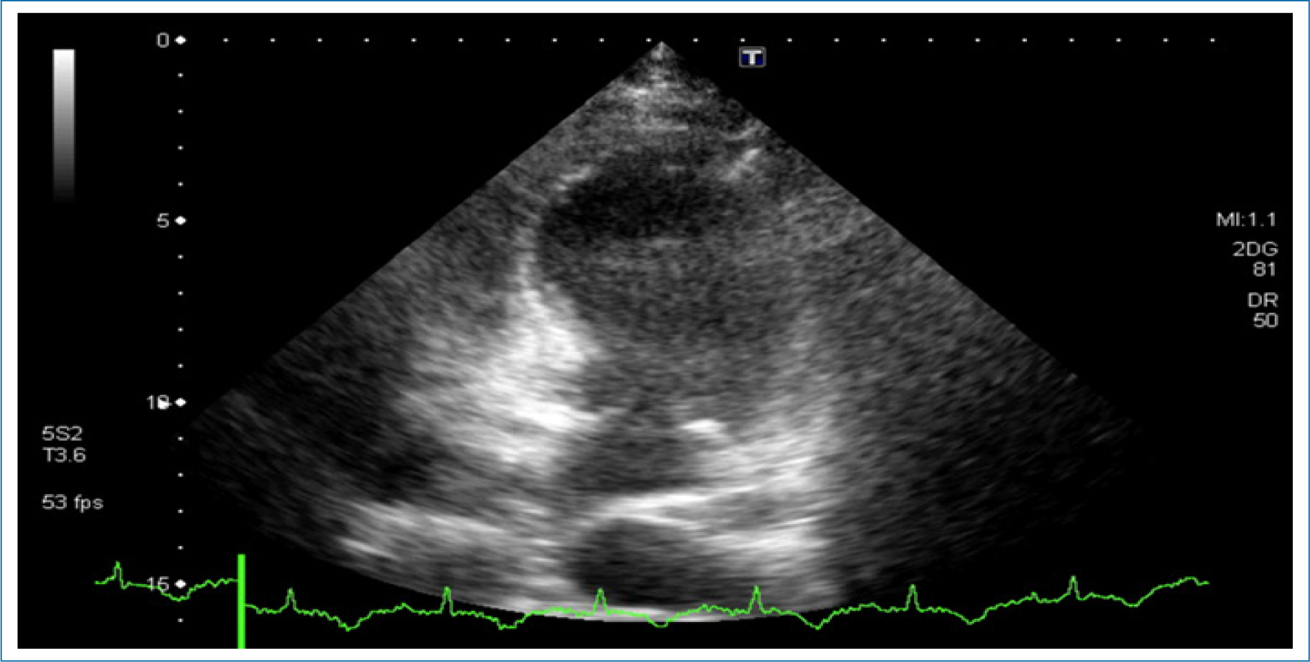
Figure 2. Transthoracic end-systolic four-chamber apical view showing a non-dilated left ventricle with apical and midventricular akinesis and hyperkinesis of the basal segments. Left ventricular systolic function was severely impaired, while the right ventricle size and function remained normal.
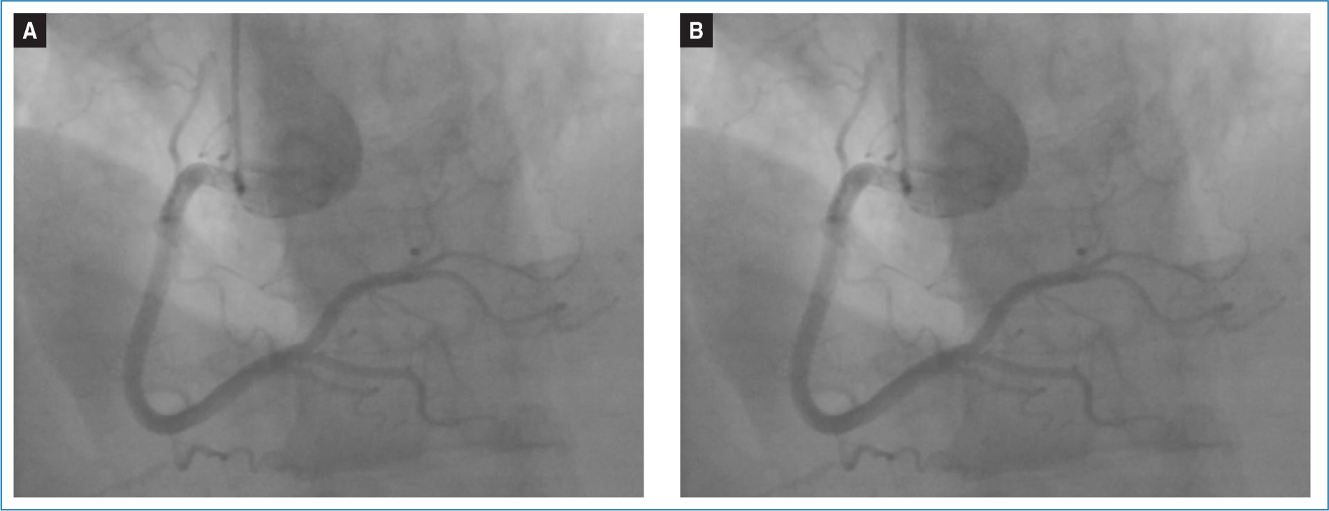
Figure 3. Coronary angiogram showing A: left and B: right coronary artery selective contrast infusions without significant obstructions.
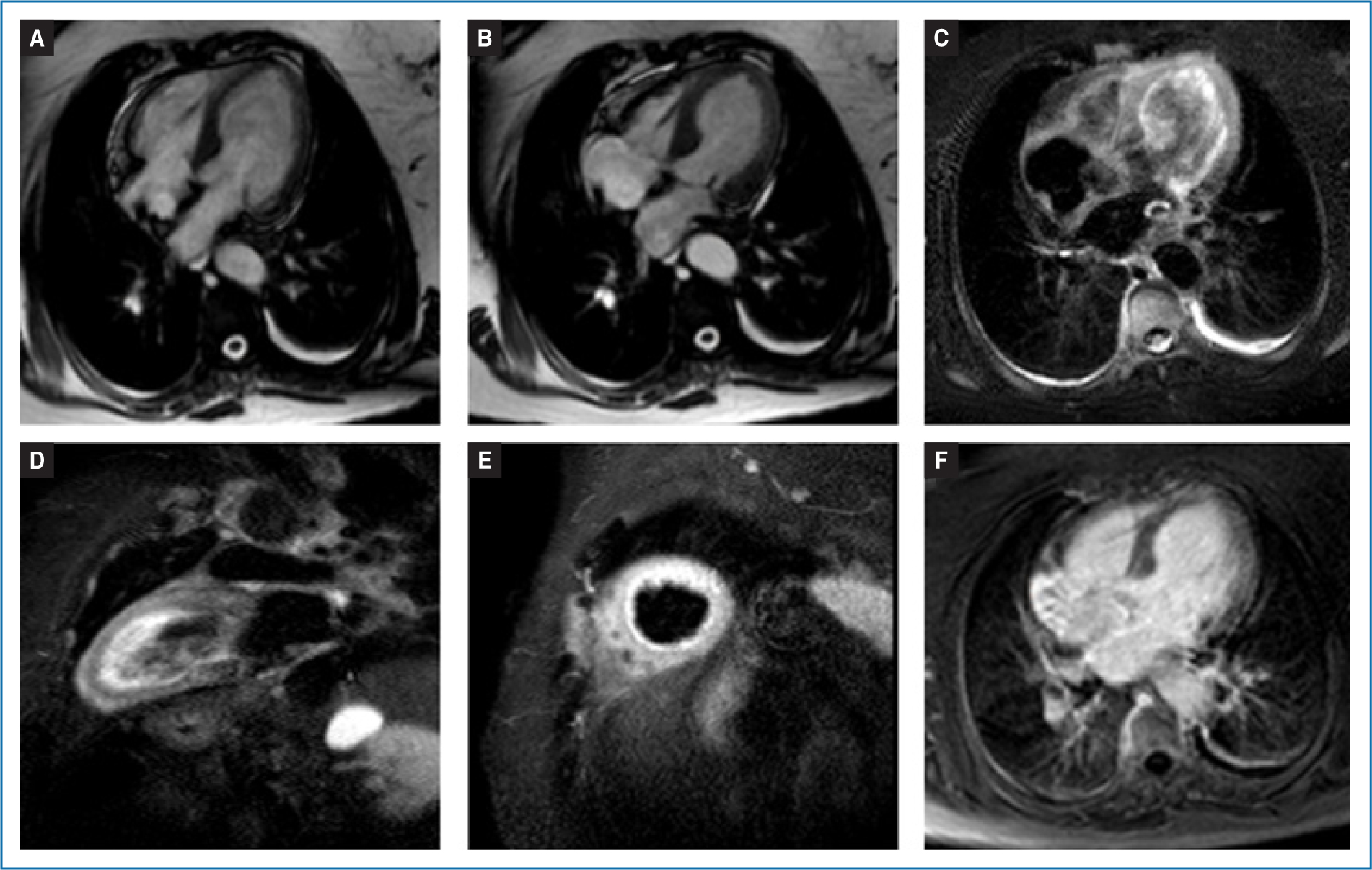
Figure 4. Cardiac magnetic resonance (MR) imaging performed on admission. Four-chamber cardiac MR cine frame in end-diastole and end-systole A and B: showing circumferential hypokinesia of apical and midventricular segments. C: T2-weighted short inversion time inversion recovery images of four-chamber and D and E: the long and short 2 chamber views, showing diffuse increase of myocardial signal (edema) in the hypokinetic segments. F: No late gadolinium enhancement was detected in the affected segments in the gadolinium-enhanced T1-weighted MR sequences.
Once heart failure medication (nebivolol 2.5 mg o.i.d, candesartan 4 mg o.i.d; spironolactone 25 mg o.i.d and furosemide 40 mg o.i.d) was titrated, the ventricular systolic function, the abnormalities of the EKG (Fig. 5), and the cardiac biomarkers progressively normalized. At 2 weeks from hospital discharge, LVEF had normalized to 61%, with complete resolution of the wall motion anomalies. After 4 weeks, under close follow-up in the cardio-oncology clinic, half dose of alectinib was restarted (300 mg o.i.d) and slowly titrated up to 600 mg b.i.d in a weekly-basis, without clinical, analytical, EKG, or echocardiographic evidence of TTS recurrence. Due to tumor progression in February 2020, alectinib was discontinued, and fourth-line ceritinib (450 mg o.i.d taken with food) was made initiated with overall good tolerance and partial response achieved. Nowadays, the patient remains on treatment with lorlatinib, originally initiated at 100mg o.i.d., since her last tumor progression in June 2024.
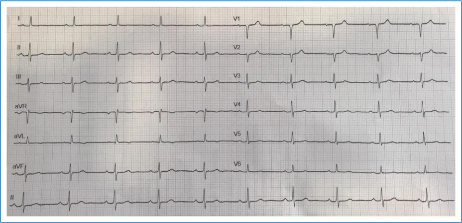
Figure 5. ECG on discharge showing resolution of the previous repolarization abnormalities.
Discussion
This clinical case represents the first documented TTS directly related to alectinib treatment. Although isolated cases have been reported in pharmacovigilance sources,11 typical TTS in a patient under recent initiation of alectinib has not been previously documented. TTS represents a rare expression of cancer treatment-related cardiac disfunction with no unequivocal predisposing factors.7 However, in the reported case, the condition developed in the context of the classical phenotype, a postmenopausal woman exposed to a physical stressor, which in this case was alectinib. The underlying pathophysiology is not completely understood, but it is characteristically elicited by an emotional or physical stressor where an endogenous release of catecholamine leads to subsequent coronary endothelial dysfunction.7,12 Patients with a history of malignancy exhibit a greater risk of developing TTS, and cancer therapies reported to trigger TTS include 5-flourouracil, capecitabine, rituximab, and immune checkpoint inhibitors.9 What other mechanisms can underlie after alectinib initiation may need further investigation.1–3
Alectinib reported cardiotoxicity is rare, with sinus bradycardia and a mild increase of QT interval (mean change of 5.3 ms) being the most commonly reported cardiac adverse effects. Grade 1 or 2 bradycardia (8.9%) have been reported in patients treated with alectinib in clinical trials.4,13 In the phase III ALEX clinical trial, 15% of patients treated with alectinib had heart rate values below 50 bpm compared with 20% of patients treated with crizotinib.1 No case of bradycardia led to discontinuation of alectinib. A wide time lag has been reported after therapy initiation, usually between 26 h and up to 3 months.14 In previous reports, crizotinib and alectinib were more likely to cause arrhythmia, while crizotinib and lorlatinib were more likely to cause blood pressure abnormalities.15 Nevertheless, although the incidence of bradycardia was initially reported as < 10%, recent studies such as Pruis et al. reported a 42% incidence, being mostly asymptomatic.4
The present case contributes additional clinical and imaging data, reinforcing the need for increased awareness of stress-induced cardiomyopathy in patients treated with alectinib. Limitations include causality cannot be definitively established in a single case report, and alternative contributing factors such as cancer-related stress cannot be completely excluded. In addition, long-term follow-up beyond the acute episode is limited, and pharmacovigilance reporting data are scarce, which limits broader generalization.
Further investigation is needed to determine whether this association is coincidental or represents a true adverse effect, as well as to identify possible predisposing factors.
Conclusion
The occurrence of cardiac symptoms after initiation of cancer treatment requires an evaluation with ECG, biomarkers, and cardiac imaging to rule out cancer treatment-related cardiac disfunction. TTS should be considered in patients receiving alectinib who present with acute cardiac dysfunction, but alternative causes must always be carefully excluded. If a potential association with the drug is suspected, the event should be reported to the Spanish Pharmacovigilance System (AEMPS). Since physiopathogenic mechanisms of alectinib-related TTS are unknown, rechallenge of alectinib should be approached with caution, under close surveillance in collaboration with a cardio-oncology team to ensure patient safety without compromising tumor prognosis.
Funding
None.
Conflicts of interest
None.
Ethical considerations
Protection of human subjects and animals. The authors declare that no experiments on humans or animals were performed for this research.
Confidentiality, informed consent, and ethical approval. The authors have obtained approval from the Ethics Committee for the analysis of routinely collected and anonymized clinical data; therefore, individual informed consent was not required. Relevant ethical recommendations have been followed.
Declaration on the use of artificial intelligence. The authors declare that no generative artificial intelligence was used in the writing or creation of the content of this manuscript.
